What Is Electromyography?
Electromyography (EMG) is a diagnostic test that is commonly used by physicians who want to evaluate the health of muscle tissue as well as the motor neurons (nerve cells) that regulate muscle activity. In particular, if it is believed that a certain spinal condition is disrupting the signal transmission from nerves within the spinal cord that are responsible for controlling muscles then electromyography will be performed.
Specific nerve cells called motor neurons transmit signals that cause muscles to stretch and contract. An electromyography records the signals and converts them into information that is displayed on a monitor. The information can then be interpreted by a specialist, usually a neurologist, as this type of physician specializes in the brain, spinal cord, and nerve disorders. However, a highly-trained technician may also perform the procedure. The results of electromyography can reveal muscle or nerve dysfunction as well as problems with the transmission of signals from the motor neurons to the muscles.
How Is Electromyography Performed?
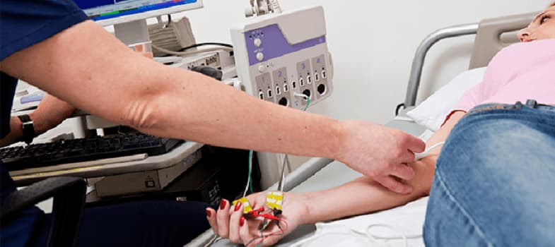
Electromyography involves inserting one or more thin needle electrodes directly into a patient’s muscle tissue in order to record its electrical activity. A ground electrode is also placed under either the arm or the leg. In some cases, up to five or more needles may need to be inserted in order to conduct the test. After the electrodes are in place, the patient may be requested to engage in movements that cause the muscles to slightly contract and forcefully contract, but measurements are also made while the muscles are at rest. Electrical signals are not produced when muscle tissue is at rest and this helps ensure that the electromyograph is not producing false readings.
The electrodes transmit the electrical activity in the form of waves to an oscilloscope, which is the monitor that the recorded activity is displayed on. The activity can also be heard through the use of an audio-amplifier. The shape and size of the waves that are observed on the oscilloscope provide the specialist with information regarding the muscle’s ability to react when it is stimulated by nerves. The more forceful the muscle contractions, the more waves that will be observed on the oscilloscope.
Electromyography is an outpatient procedure that generally does not pose any risks or side effects. However, due to the use of needles, there is a small risk of developing an infection, bleeding, or nerve injury at the site where the electrodes are inserted. Certain lifestyle habits can influence the test results so in some cases, patients are asked to refrain from drinking caffeinated beverages such as sodas, coffee, or tea and smoking cigarettes for at least two hours before undergoing electromyography. Medication, both over-the-counter and prescription, as well as herbal supplements can also alter the results so patients are usually asked about medication or supplements they may be taking.

Conditions Related To Electromyography
Electromyography is often ordered if a patient is displaying symptoms that indicate the presence of a muscle or nerve disorder. These symptoms include:
- Muscle weakness
- Muscle cramping and pain
- Numbness
- Tingling
- Sharp, stabbing pain in the limbs
Conditions that may be diagnosed or ruled out based on electromyography results include:
- Polymyositis or muscular dystrophy, which are muscle disorders
- Myasthenia gravis, which is a disease the affects the connections between nerves and muscles
- Carpal tunnel syndrome or peripheral neuropathies, which refers to damaged nerves
- Amyotrophic lateral sclerosis or polio, which are disorders that affect motor neurons in the spinal cord
- A herniated disc, which is a condition that affects nerve roots
Electromyography may also be performed during procedures, such as spinal surgery for a herniated disc, in order to monitor spinal cord activity and prevent unnecessary complications.
Conclusions
Electromyography is a diagnostic test that physicians generally order if they want to evaluate the health of muscle tissue and specific nerve cells called motor neurons that regulate muscle movement. This test is most often used to determine whether a spinal condition may be hindering the transmission of signals from nerves to the muscles. Active muscles produce electrical signals that can be recorded by thin needle electrodes and transmitted to a monitor that displays the signals as waves. The size and shape of the waves indicate how well muscles are responding to the nerves that regulate them. Electromyography is an outpatient procedure and it generally does not cause complications.
Muscle weakness, cramping, pain, numbness, tingling, and sharp stabbing pain in the extremities usually indicate that a muscle or nerve disorder may be present. A specialist such as a neurologist or highly-trained technician can perform electromyography to determine what type of condition may be causing a patient’s symptoms. Common conditions that may be diagnosed through this test include muscular dystrophy, polymyositis, carpal tunnel syndrome, amyotrophic lateral sclerosis, or a herniated disc. Although electromyography can be used to diagnose numerous muscle and nerve disorders, it can also be performed during surgeries (e.g., spinal surgery) to monitor spinal cord and nerve activity.
References
- Ghosh PS, Sorenson EJ. Diagnostic yield of electromyography in children with myopathic disorders. Pediatr Neurol. 2014; 51(2):215-9.
- Derry KL, Venance SL, Doherty TJ. Decomposition-based quantitative electromyography in the evaluation of muscular dystrophy severity. Muscle Nerve. 2012; 45(4):507-13.
- Somnier FE, Skeie GO, Aarli JA, Trojaborg W. EMG evidence of myopathy and the occurrence of titin autoantibodies in patients with myasthenia gravis. Eur J Neurol. 1999; 6(5):555-63.
- Wee AS. Needle electromyography in carpal tunnel syndrome. Electromyogr Clin Neurophysiol. 2002; 42(4):253-6.
- Krarup C. Lower motor neuron involvement examined by quantitative electromyography in amyotrophic lateral sclerosis. Clin Neurophysiol. 2011; 122(2):414-22.
- Fotakopoulos G, Alexiou GA, Pachatouridis D, Karagiorgiadis D, Konitsiotis S, Kyritsis AP, Voulgaris S. The value of transcranial motor-evoked potentials and free-running electromyography in surgery for cervical disc herniation. J Clin Neurosci. 2013; 20(2):263-6.
- Nichols GS, Manafov E. Utility of electromyography for nerve root monitoring during spinal surgery. J Clin Neurophysiol. 2012; 29(2):140-8.